
By Håkan Boström, MD, Senior Consultant and Pediatric Radiologist, Liz Ivarsson, MD, Senior Consultant and Pediatric Radiologist, and Pär-Arne Svensson, BSc, Research Radiographer MRI, Queen Silvia Children’s Hospital, Gothenburg, Sweden
Tuberous sclerosis complex (TSC) is a genetic disorder that causes the formation of benign tumors, or tubers, primarily in the brain, heart, kidneys, eyes, skin and lungs. In the brain, there is a risk for development of subependymal giant cell astrocytoma (SEGA) and in the kidneys a risk for development of angiomyolipoma (AML) and/or renal cell carcinoma. Seizures, cognitive difficulties and behavioral or developmental disorders are also hallmarks of TSC.
The growth of SEGA can potentially block the flow of cerebrospinal fluid within the brain, causing a buildup of fluid and pressure that can lead to headaches and blurred vision. Although AML typically does not have any symptoms, it can cause pain and in some instances lead to anemia or a life-threatening hemorrhage.
Due to the potential complications resulting from TSC, these patients undergo annual brain and kidney examinations to check for the presence of SEGA in the brain and cysts and/or AML in the kidneys. High image quality and short scan time are desired in these pediatric imaging cases.
Patient history
A 16-year-old patient weighing 68 kg (150 lb) with TSC, epilepsy, developmental disability and autism underwent MR imaging for an annual brain and body (kidney) examination to check for SEGA in the brain and cysts or AML in the kidneys.
Results
In the brain, no change or sign of SEGA was detected from the prior exam (one year earlier). Multiple small cortical tubers in both the frontal lobes and left parietal lobe were noted.
In the left kidney, a new atypical fat-poor AML in the lower pole was detected.
Discussion
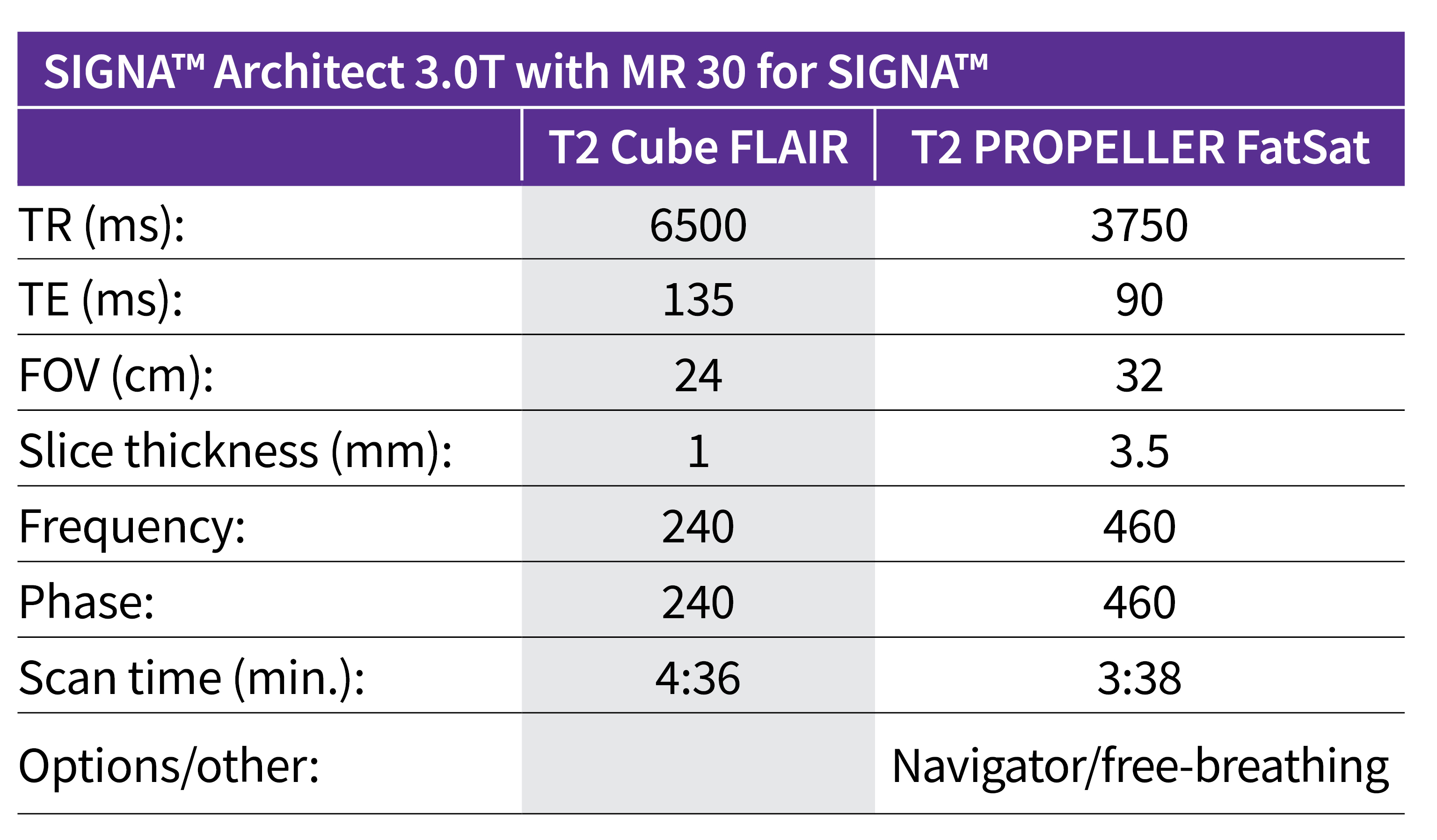
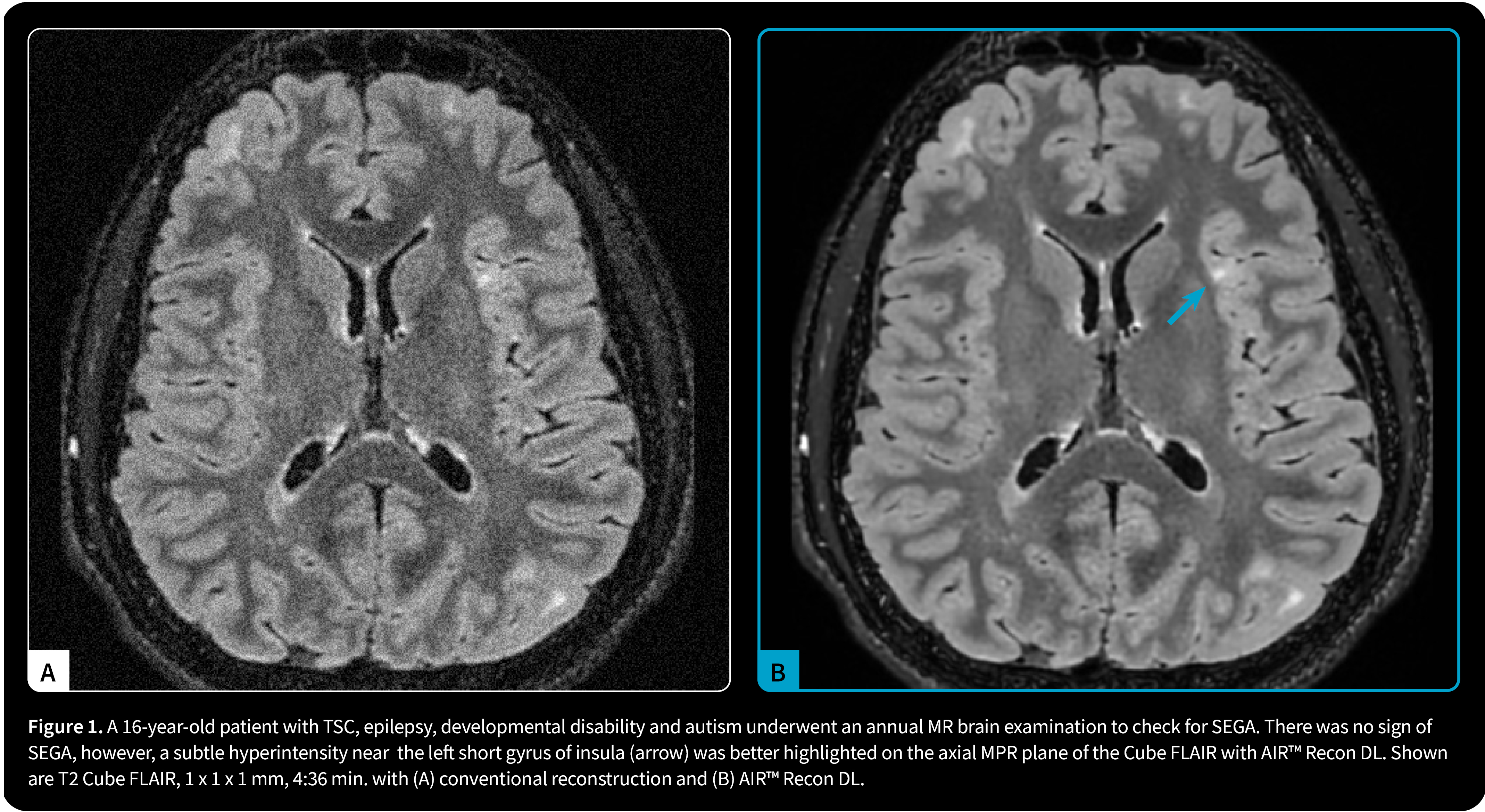
The addition of AIR™ Recon DL to both 3D and PROPELLER sequences in MR 30 for SIGNA™ led to image quality improvement, as well as reduced scan time. Shorter scan times are particularly important in pediatric imaging, especially when imaging patients with developmental or behavioral disorders who may not be able to remain still during the MR examination.
Although there was no sign of SEGA, subtle subcortical hyperintensities, for example one near the left short gyrus of insula, were better highlighted on the axial MPR plane of the Cube FLAIR with AIR™ Recon DL. The significant reduction in Gibbs artifacts and the increase in SNR with AIR™ Recon DL made the visualization of subtle hyperintensities easier. Also, the gain in SNR enabled the acquisition of a 1 mm isotropic acquisition, which made the reconstructed plane as informative as the initially acquired plane.
By utilizing AIR™ Recon DL PROPELLER, it was possible to acquire the kidney with free-breathing and exceptional image quality. PROPELLER is designed to reduce the effect of patient voluntary and physiologic motion (breathing, flow, peristalsis), enabling consistently good, diagnostic-quality images even for challenging patients and difficult-to-image anatomy. The combination of FatSat, PROPELLER and AIR™ Recon DL allowed for the detection of small abnormalities like the one in the right kidney with confidence.
Read the latest issue of GE HealthCares MR "SIGNA™ Pulse Magazine"